

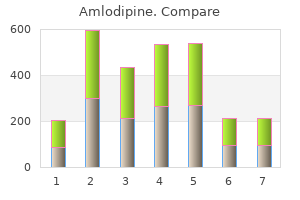
"Generic 5mg amlodipine, arteria inominada".
By: S. Tom, M.A., Ph.D.
Clinical Director, Medical College of Georgia at Augusta University
With types 1 and 2 this can usually be done closed; the part is then splinted securely for 36 weeks arrhythmia echocardiogram generic amlodipine 5mg visa. An attempt can be made to achieve this by gentle manipulation under general anaesthesia; if this is successful blood pressure high symptoms discount amlodipine 5 mg mastercard, the limb is held in a cast for 48 weeks (the longer periods for type 4 injuries) arteriogram definition buy genuine amlodipine. If a type 3 or 4 fracture cannot be reduced accurately by closed manipulation blood pressure chart symptoms buy amlodipine 5mg online, immediate open reduction and internal fixation with smooth K-wires is essential. The limb is then splinted for 46 weeks, but it takes that long again before the child is ready to resume unrestricted activities. The fracture does not traverse the width of the physis; after reduction (b) bone growth is not distorted. Complications Types 1 and 2 injuries, if properly reduced, have an excellent prognosis and bone growth is not adversely affected. Exceptions to this rule are injuries around the knee involving the distal femoral or proximal tibial physis; both growth plates are undulating in shape, so a transverse fracture plane may actually pass through more than just the hypertrophic zone but also damage the proliferative zone. Complications such as malunion or non-union may also occur if the diagnosis is missed and the fracture remains unreduced. Types 3 and 4 injuries may result in premature fusion of part of the growth plate or asymmetrical growth of the bone end. If the bridge is relatively small (less than one-third the width of the physis) it can be excised and replaced by a fat graft, with some prospect of preventing or diminishing the growth disturbance (Langenskiold, 1975; 1981). Established deformity, whether from asymmetrical growth or from malunion of a displaced fracture. The injury is one in which the joint is momentarily twisted or bent into an abnormal position. Tenderness is localized to the injured ligament and tensing the tissues on that side causes a sharp increase in pain. If the force is great enough the ligaments may tear, or the bone to which they are attached may be pulled apart. The articular cartilage, too, may be damaged if the joint surfaces are compressed or if there is a fracture into the joint. As a general principle, forceful angulation will tear the ligaments rather than crush the bone, but in older people with porotic bone the ligaments may hold and the bone on the opposite side of the joint is crushed instead, while in children there may be a fractureseparation of the physis. Treatment the joint should be firmly strapped and rested until the acute pain subsides. Thereafter, active movements are encouraged, and exercises practised to strengthen the muscles. Sometimes the ligament holds and the bone to which it is attached is avulsed; this is effectively the same lesion but easier to deal with because the bone fragment can be securely reattached. As with a strain, the joint is suddenly forced into an abnormal position; sometimes the patient actually hears a snap. The joints most likely to be affected are the ones that are insecure by virtue of their shape or least well protected by surrounding muscles: the knee, ankle and finger joints. Pain is severe and there may be considerable bleeding under the skin; if the joint is swollen, this is probably due to a haemarthrosis. The patient is unlikely to permit a searching examination, but under general anaesthesia the instability can be demonstrated; it is this that distinguishes the lesion from a strain. Strictly speaking, a sprain is any painful wrenching (twisting or pulling) movement of a joint, but the term is generally reserved for joint injuries less severe than actual tearing of the capsule or ligaments. If the stretching or twisting force is severe enough, the 730 23 Principles of fractures (a) (b) (c) (d) (e) 23. If the soft tissues hold, the bone on the opposite side may be crushed (b), or a fragment may be pulled off by the taut ligament (c). Subluxation (d) means the articular surfaces are partially displaced; dislocation (e) refers to complete displacement of the joint. Clinical features Following an injury the joint is painful and the patient tries at all costs to avoid moving it. The limb is often held in a characteristic position; movement is painful and restricted.
Aftercare In the ward blood pressure medication effect on running purchase amlodipine 2.5mg on-line, the limb is elevated and its circulation carefully watched blood pressure of 10060 discount amlodipine express. Antibiotic cover is continued but only for a maximum of 72 hours in the more severe grades of injury heart attack 60 amlodipine 2.5mg on-line. Wound cultures are seldom helpful as osteomyelitis hypertension kidney specialists lancaster pa purchase amlodipine online from canada, if it were to ensue, is often caused by hospital-derived organisms; this emphasizes the need for good debridement and early fracture cover. On resolution of the infection, attention can be given to stabilizing the fracture so that joint movement can recommence. Permanent stiffness is a real threat; where fracture stabilization cannot be achieved to allow movement, the joint should be splinted in the optimum position for ankylosis, lest this should occur. Tissue damage is produced by: (1) direct injury in the immediate path of the missile; (2) contusion of muscles around the missile track and (3) bruising and congestion of soft tissues at a greater distance from the primary track. With high-velocity missiles (bullets, usually from rifles, travelling at speeds above 600 m/s) there is marked cavitation and tissue destruction over a wide area. The splintering of bone resulting from the transfer of large quantities of energy creates secondary missiles, causing greater damage. With low-velocity missiles (bullets from civilian hand-guns travelling at speeds of 300600 m/s) cavitation is much less, and with smaller weapons tissue damage may be virtually confined to the bullet track. However, with all gunshot injuries debris is sucked into the wound, which is therefore contaminated from the outset. If the injury is to soft tissues only with minimal bone splinters, the wound may be safely treated without surgery but with local wound care and antibiotics. The method of wound closure will depend on the state of tissues after several days; in some cases delayed primary suture is possible but, as with other open injuries, close collaboration between plastic and orthopaedic surgeons is needed (Dicpinigaitis et al. Close-range shotgun injuries, although the missiles may be technically low velocity, are treated as highvelocity wounds because the mass of shot transfers large quantities of energy to the tissues. The wounds should each be covered with a sterile dressing and the area examined for artery or nerve damage. Antibiotics should be given immediately, following the recommendations for open fractures (see Table 23. Local complications can be divided into early (those that arise during the first few weeks following injury) and late. Definitive treatment Traditionally, all missile injuries were treated as severe open injuries, by exploration of the missile track and formal debridement. Even if its outward appearance is normal, the intima may be detached and the vessel blocked by thrombus, or a segment of artery may be in spasm. The effects vary from transient diminution of blood flow to profound ischaemia, tissue death and peripheral gangrene. Clinical features the patient may complain of paraesthesia or numbness in the toes or the fingers. The injured limb is cold and pale, or slightly cyanosed, and the pulse is weak or absent. If a vascular injury is suspected an angiogram should be performed immediately; if it is positive, emergency treatment must be started without further delay. The fracture is re-x-rayed and, if the position of the bones suggests that the artery is being compressed or kinked, prompt reduction is necessary. If there is no improvement, the vessels must be explored by operation preferably with the benefit of preoperative or peroperative angiography. A cut vessel can be sutured, or a segment may be replaced by a vein graft; if it is thrombosed, endarterectomy may restore the blood flow. If vessel repair is undertaken, stable fixation is a must and where it is practicable, the fracture should be fixed internally. The plain x-ray (a) looked as if the proximal bone fragment might have speared the popliteal artery. Bleeding, oedema or inflammation (infection) may increase the pressure within one of the osseofascial compartments; there is reduced capillary flow, which results in muscle ischaemia, further oedema, still greater pressure and yet more profound ischaemia a vicious circle that ends, after 12 hours or less, in necrosis of nerve and muscle within the compartment. A similar cascade of events may be caused by swelling of a limb inside a tight plaster cast. The telltale signs should be looked for (and documented) during the initial examination and again after reduction of the fracture. Closed nerve injuries In closed injuries the nerve is seldom severed, and spontaneous recovery should be awaited it occurs in 90 per cent within 4 months. Clinical features High-risk injuries are fractures of the elbow, forearm bones, proximal third of the tibia, and also multiple Open nerve injuries With open fractures the nerve injury is more likely to be complete.
Amlodipine 10mg mastercard. Is Olive Oil Healthy? | Dr. Josh Axe.
If bone is exposed and length of the digit is important for the individual patient blood pressure medication ratings buy generic amlodipine pills, then an advancement flap or neurovascular island flap should be considered hypertension signs cheap generic amlodipine uk. In young children heart attack 50 damage discount amlodipine online amex, the finger-tips recover extraordinarily well from injury and they should be treated with dressings rather than grafts or terminalization arrhythmia during pregnancy cheap generic amlodipine uk. Thumb length should never be sacrificed lightly and every effort should be made to provide a long, sensate digit. Nail bed injuries Nail bed injuries are often seen in association with fractures of the terminal phalanx. If appearance is important, meticulous repair of the nail bed under magnification, replacing any loss with a split thickness nail bed graft from one of the toes, will give the best cosmetic result. In full thickness wounds without bone exposure, the wound should be thoroughly cleaned and then covered with a nonadherent dressing. A light plaster slab holds the wrist and hand in the position of safety (wrist extended, metacarpo-phalangeal joints flexed to 90 degrees, interphalangeal joints straight, thumb abducted). This is the position in which the metacarpo-phalangeal and interphalangeal ligaments are fully stretched and fibrosis therefore least likely to cause contractures. Failure to appreciate this point is the commonest cause of irrecoverable stiffness after injury (see Fig 16. There should be minimal restriction at the front of the fingers, otherwise the resistance can precipitate rupture of the tendon. Various protocols are followed for flexor tendon injuries, including passive, active or elastic-band assisted flexion. In all cases the risk of rupture is balanced against the need for early mobilization. If secondary surgery is required, tendon or nerve repair is postponed until the skin is healthy, there is no oedema and the joints have regained a normal range of passive movement. Replantation With modern microsurgical techniques and appropriate skill, amputated digits or hands can be replanted. An amputated part should be wrapped in sterile saline gauze and placed in a plastic bag, which is itself placed in watery ice. For a hand or forearm, the cold ischaemic time is only about 12 hours and the warm time much less. After resuscitation and attention to other potentially life-threatening injuries, the patient and the amputated part should be transferred to a centre where the appropriate surgical skills and facilities are available. Furthermore, and perhaps most importantly, it depends on whether the replanted part is likely to give better function than an amputation. If the latter is used, the sling must be removed several times a day to exercise the elbow and shoulder. Splintage should allow as many joints as possible to be exercised, consistent with protecting the repair. Proximal amputations (through the palm, wrist or forearm) likewise merit an attempt at replantation. There is a high complication rate, including stiffness, non-union, poor sensation, and cold intolerance; a replanted single finger is likely to be excluded from use. The exception is an amputation beyond the insertion of flexor digitorum superficialis, when a cosmetic, functioning finger-tip can be retrieved. Severely crushed, mangled or avulsed parts may not be replantable; and parts with a long ischaemic time may not survive. General medical disorders or other injuries may engender unacceptable risks from the prolonged anaesthesia needed for replantation. Superficial burns are covered with moist non-adherent dressings; the hand is elevated and finger movements are encouraged. Partial thickness burns can usually be allowed to heal spontaneously; the hand is dressed with an antimicrobial cream and splinted in the position of safety. Devitalized tissue should be excised; the wound is cleaned and dressed and 25 days later skin-grafted.
The cerebrum arterial nicking quality 10mg amlodipine, the largest part of the brain hypertension nos discount amlodipine american express, is responsible for receiving and processing stimuli arteria inominada order amlodipine american express, initiating voluntary movement hypertension 99791 best 5 mg amlodipine, and storing information. The cerebral cortex (outer region) is made up of gray matter and is arranged in folds (Figure 139). The brain also has small cavities called ventricles (vehntrih-kuhlz) (Figure 1310). There are four ventricles of the brain: two lateral ventricles, a third ventricle, and a fourth ventricle. The ventricles of the brain (and central canal of the spinal cord) are lined with a membrane called the ependyma (eh-pehn-dih-mah). The cerebellum is the second largest part of the brain, and it coordinates muscle activity for smooth movement. The cerebellum has an inner portion, called the vermis (vr-mihs) because it is wormlike, and other portions divided into right and left cerebellar hemispheres. The brainstem is the stalklike portion of the brain that connects the cerebral hemispheres with the spinal cord. The interbrain contains structures such as the pituitary gland, hypothalamus, and thalamus. These structures are responsible for endocrine activity, regulation of thirst and water balance, and regulation of body temperature. The midbrain contains structures responsible for Sulcus Gyrus Hemisphere Hemisphere Hemisphere Fissure Cerebellum Frontal Parietal Temporal Occipital Figure 139 Dorsal view of the cerebral cortex. Each hemisphere is further divided into lobes, and each lobe is named for the bone plate covering it: frontal (frohn-tahl) lobe = most cranial lobe that controls motor function; parietal (pahr-ih-tahl) lobe = receives and interprets sensory nerve impulses; occipital (ohcks-ihp-ih-tahl) lobe = most caudal lobe that controls vision; temporal (tehmp-ruhl) lobe = laterally located lobe that controls hearing and smell. Nerves of Steel Lateral ventricle (2) Fourth ventricle (1) 271 Some parts of the brain are named not based on location or division, but rather on how they look. Another example is the hippocampus, a portion of the limbic system that involves memory. The hippocampus (hihp-kahm-puhs) is shaped like a seahorse, which exists in mythology as a sea monster with the head of a horse and the tail of a fish and as an actual sea creature. The name hippocampus comes from the Greek hippos, meaning horse, and kampos, meaning sea monster. The arbor vitae (tree of life) of the cerebellum are the treelike outlines seen on sagittal views of the cerebellum. The pons (pohnz) is the bridge at the base of the brain that allows nerves to cross over so that one side of the brain controls the opposite side of the body. The medulla oblongata (meh-duhl-ah ohb-lohng-gahtah) is the cranial continuation of the spinal cord that controls basic life functions. The spinal cord passes through an opening in the occipital bone called the foramen magnum (fr-mehn mahg-nuhm). The spinal cord carries all of the tracts that influence the innervation of the limbs and lower part of the body. In addition, the spinal cord is the pathway for impulses going to and from the brain. The gray matter of the spinal cord is located in the internal portion and is not protected by myelin. The white matter of the spinal cord is located in the external portion and is myelinated. The medical term for swelling (normal or abnormal) is intumescence (ihn-too-meh-sehns). The swelling is caused by an increase in white matter and cell bodies that are associated with the innervation of the limbs. One swelling occurs in the area of C6T2, which is known as the cervical intumescence. The other swelling occurs in the area of the L4caudal segment, which is known as the lumbosacral intumescence. The cranial parts of the spinal cord have tracts with fibers from the cranial and caudal portions, but the caudal parts of the spinal cord have only tracts with fibers from the caudal portions. Therefore, as the spinal cord proceeds caudally, its cross-sectional area decreases. At the level of the cranial lumbar vertebrae, the spinal cord becomes cone-shaped.